


Highlights of this edition:
Small cell lung cancer (SCLC) organoids from non-invasive sources reveal drug target
A fully biological organ-on-a-chip fabricated using bioprinting
Full database of latest scientific paper on organoids with “Organoids Digest Extended”
More details below 👇
Welcome to Organoids Digest, your bi-weekly update on organoids, organ-on-a-chip, and 3D cell culture.
In this issue, we explore how precise bioprinting and laser sculpting techniques are redefining what's possible in organoid research. We also highlight exciting progress on a promising new therapeutic target for small-cell lung cancer (SCLC).
We wish you an enjoyable read ❤️
Article: Engineered epithelial curvature controls Paneth cell localization in intestinal organoids
F. Max Yavitt et al | April 22, 2025 | Cell Biomaterials
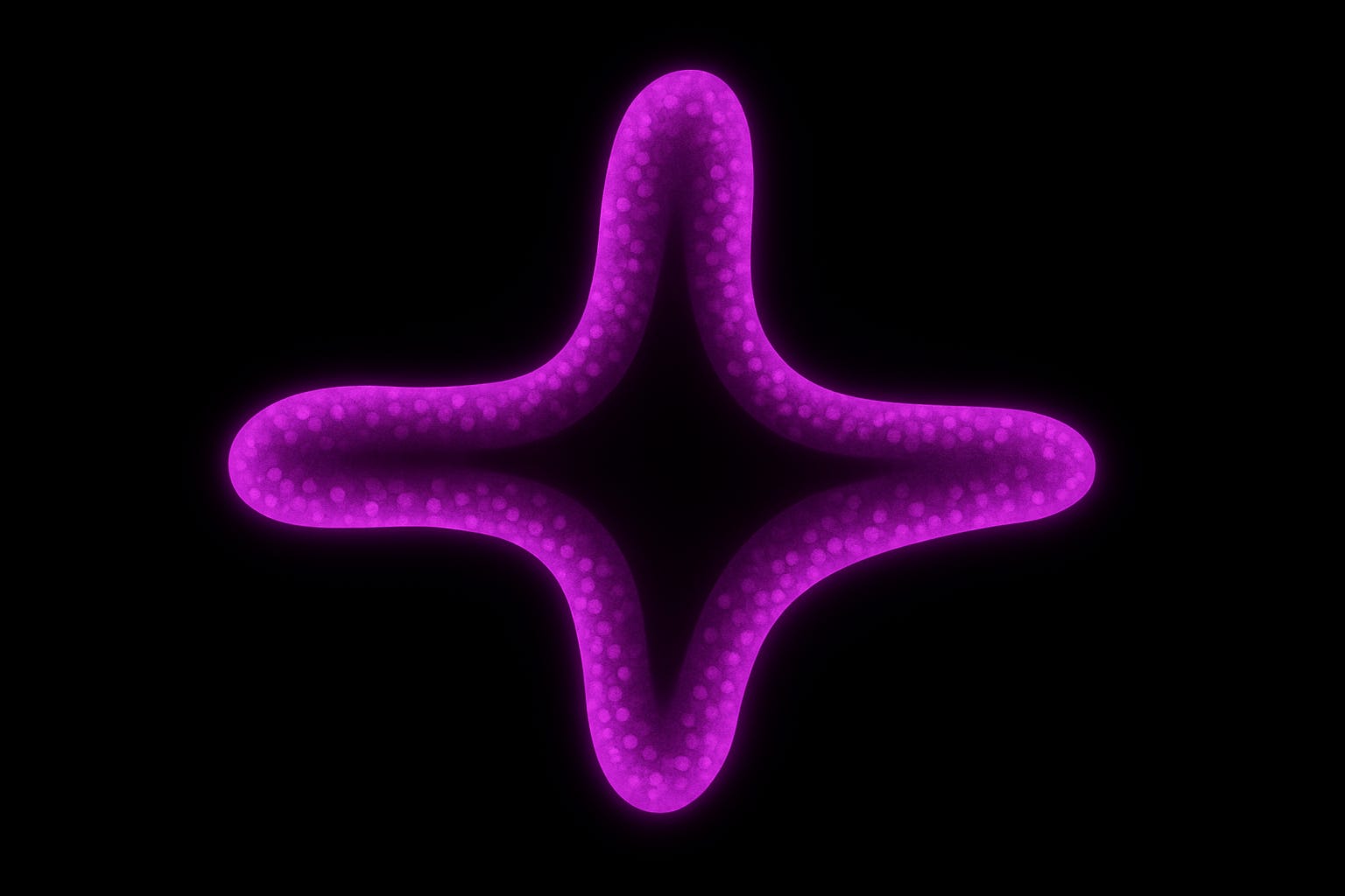
Designer crypts: Shaping organoids by laser precision
Since the landmark report from Hans Clevers’ lab that intestinal stem cells can self-organise into crypt-bearing “mini-guts”, researchers have pushed to make them not just 3D, but functionally and structurally accurate.
A key challenge? In traditional Matrigel-based systems, organoids naturally form crypts with stem cells and Paneth cells (PCs), but these crypts have highly variable shapes, and PCs scatter unpredictably; a significant limitation when crypt architecture itself critically modulates stem-cell behavior.
This new study offers a solution: swap out variable Matrigel for a synthetic, photodegradable PEG hydrogel, then sculpt it with a laser to create crypts of precise curvature.
The method:
Organoids are grown in a PEG matrix whose stiffness is locally reduced by a confocal laser, carving narrow (30–60 µm) or trapezoidal channels around them.
These pre-patterned “crypt traps” guide epithelial growth into predictable shapes.
Live imaging and fluorescent PC reporters track how Paneth cells organize over time.
Key findings:
Curvature matters: Narrow, high-curvature crypts lead to earlier but fewer PC appearances. Wider, low-curvature ones attract twice as many PCs.
Extreme curvature repels PCs, while broad, shallow traps concentrate them.
These effects are independent of Matrigel: curvature alone is sufficient to dictate PC dynamics.
Why this matters: This system creates organoids with designer crypt geometry, opening up fine-tuned studies of the intestinal stem cell niche, region-specific gut functions, and disease models. It also sets the stage for applications in drug screening and host-microbiome co-culture, where precise spatial organization is key.
Resource: An organoid library unveils subtype-specific IGF-1 dependency via a YAP–AP1 axis in human small cell lung cancer
Takahiro Fukushima et al | April 30 2025 | Nature Cancer
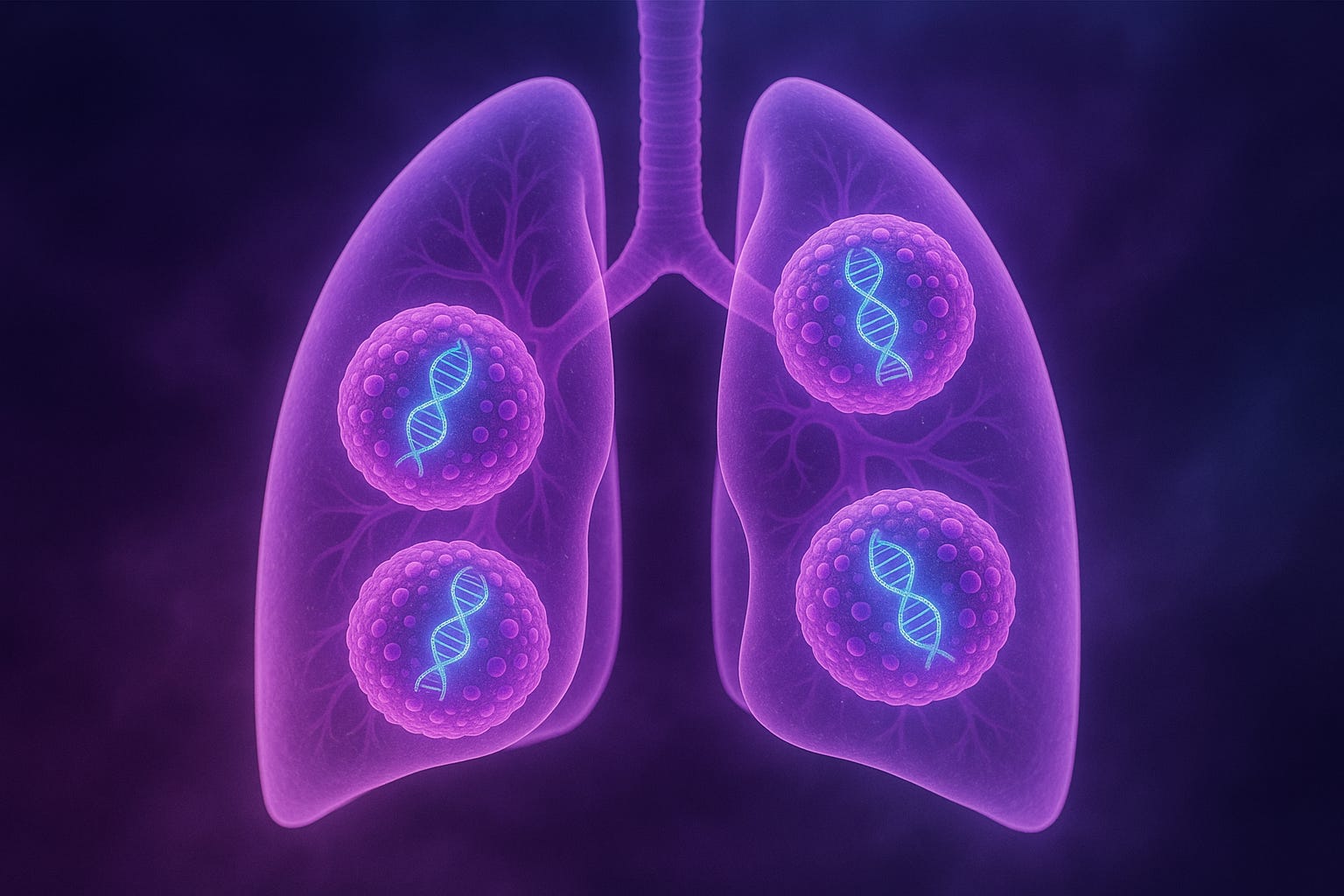
A path forward for deadly SCLC?
Small-cell lung cancer (SCLC) remains one of the deadliest cancers, with late-stage diagnoses and poor 5-year survival. But a recent study offers new hope by showing that patient-derived organoids, even from non-invasive sources like sputum and circulating tumor cells (CTCs), recapitulate key genetic alterations and transcriptional subtypes found in the disease.
What they built:
Created a 40-line SCLC organoid library, capturing all four recognized subtypes (ASCL1, NEUROD1, YAP1, POU2F3).
Each organoid line retained the transcriptional identity of its source tumor, confirmed by RNA-seq.
Key insight:
The YAP1/POU2F3 subgroup (non-neuroendocrine) showed a unique growth dependency:
IGF-1 activates IGF1R, branching into two signaling pathways:
→ AKT → AP-1
→ FAK → YAP1
Both pathways converge to drive tumor proliferation.
Therapeutic implications:
IGF1R inhibitors selectively killed YAP1/POU2F3 organoids and reduced tumor growth in xenografts.
Combining IGF1R inhibition with cisplatin showed synergistic effects.
ASCL1/NEUROD1 subtypes were unaffected, highlighting a subtype-specific vulnerability.
Mechanistic bonus:
Using CRISPR to delete TP53 and RB1 in normal lung organoids triggered IGF-1 dependency and activated YAP1—mimicking early tumor evolution toward this vulnerable subtype.
Why it matters:
This study validates a minimally invasive, scalable model for SCLC, links it to druggable subtype biology, and demonstrates how normal cells can acquire these dependencies. It represents an important advance in precision oncology, particularly in cancers where tissue access is limited.
Article: 3D bioprinting of collagen-based high-resolution internally perfusable scaffolds for engineering fully biologic tissue systems
Daniel J. Shiwarski et al | April 23, 2025 | Science Advances

Can bioprinting unlock true biological scaffolds?
Most organ-on-chip platforms host living cells inside a synthetic microfluidic chip, using PDMS or plastic channels that often have limitations such as stiffness, drug absorption, and resistance to cell remodeling.
This recent study highlights a fully biological organ-on-a-chip using collagen I scaffolds fabricated via FRESH bioprinting and perfused through a specialized (VAPOR) bioreactor. This method preserves the precise geometry characteristic of standard OoC devices with a natural ECM-like composition all achieved in a one-step process.
Key Features:
Precision printing: Collagen filaments are stabilized in a gelatin support bath, achieving <20 µm fidelity over centimeter spans.
Internal flow channels: Lumens down to 250 µm connect directly to barbed inlets, sustaining steady or pulsatile flow (up to ~3 dyne/cm²). Lower limit for perfusable channel diameter is ~100 µm; this is still challenged by photolithography (~ 10 µm) however, endothelial cells + pericytes may self-assemble to compensate.
Multi-material compatibility: Using a triple-extruder setup, distinct bioinks (e.g. fibronectin, VEGF, HUVEC-laden fibrin) can be layered with micrometer precision, offering matrices with specific properties in one system.
Biological Functionality:
Solute diffusion across collagen walls follows molecular size and improves under pressure gradients.
Endothelial and stromal cells self-assemble into capillary networks across the scaffold.
Mixed cell bioinks (e.g. MIN6 + HUVEC + MSC) generate vascularized mini-pancreases, with insulin secretion indices doubling under flow.
What’s particularly exciting is that this setup is open-source ready: any lab with a ~20 µm-accurate bioprinter can implement it.
CHIPS + VAPOR offers a rare combo: lithographic resolution, biologic material, and broad accessibility. A powerful platform for building the next generation of vascularized tissues and OoC models.
By the way: Check out the figures, they’re 👌
Organoids Digest Full Database
📄 Want even more recent and sorted resources? Check out the Organoids Digest Extended! It’s your go-to source for the latest scientific publications in organoids, organ-on-a-chip, and cutting-edge 3D models sorted by tissue and systems. Explore it by clicking below 👇

❤️ Thank you for reading this edition of Organoids Digest. If you enjoyed it, let us know by clicking the heart button below

🌐 Visit our site to learn more about 3D cell culture and organ-model know-how: cherrybiotech.com